
German Congress of Orthopaedics and Traumatology 2026 (DKOU 2026)
Sex-specific synovial fluid macrophage activation in late-stage bicondylar knee osteoarthritis
Text
Objectives and questions: Osteoarthritis (OA) is increasingly recognized as a disease driven by chronic low-grade inflammation within the joint microenvironment. Immune cell infiltration and activation, particularly of macrophages, are thought to critically contribute to disease progression. Biological sex is a major risk factor for OA, with differences in prevalence and disease burden, yet mechanisms remain poorly understood. This study aimed to characterize systemic and joint-resident macrophage subpopulations with a specific focus on compartment- and sex-dependent differences in knee OA.
Material and methods: In a cross-sectional cohort, peripheral blood (PB), synovial tissue (ST), and synovial fluid (SF) were collected from 64 patients with late-stage unicondylar (n=44) or bicondylar (n=20) knee OA (Kellgren–Lawrence III–IV) undergoing arthroplasty. Patients with infection, immunomodulatory medication, or a history of malignant or immune-mediated disease were excluded. Flow cytometry was used to quantify macrophages (CD45⁺CD14⁺) and classify subsets as activated (CD68⁺CD11b⁺), M1-like (HLA-DR⁺CD86⁺), or M2-like (CD206⁺CD163⁺). Frequencies were reported as percentages of CD45⁺ cells. Group comparisons used Student’s t-test or Mann–Whitney U test.
Results: Baseline characteristics (age, BMI, OA grade, CRP, leukocyte counts) did not differ significantly between sexes within either OA cohort. In unicondylar OA, macrophage profiles were comparable between females and males across PB, ST, and SF. In bicondylar OA, SF showed pronounced sex-specific differences, with females displaying higher frequencies of total macrophages (p = 0.033), activated macrophages (p = 0.036), and M1-like macrophages (p = 0.037) compared with males (Figure 1 [Fig. 1]). Sex-stratified comparisons between unicondylar and bicondylar OA revealed significant differences in total and activated macrophages in SF and only among female patients (Table 1 [Tab. 1]).
Figure 1: Sex-specific differences in macrophage subtypes in late-stage bicondylar knee OA (mean values ± standard deviation). Statistical significance: * p < 0.05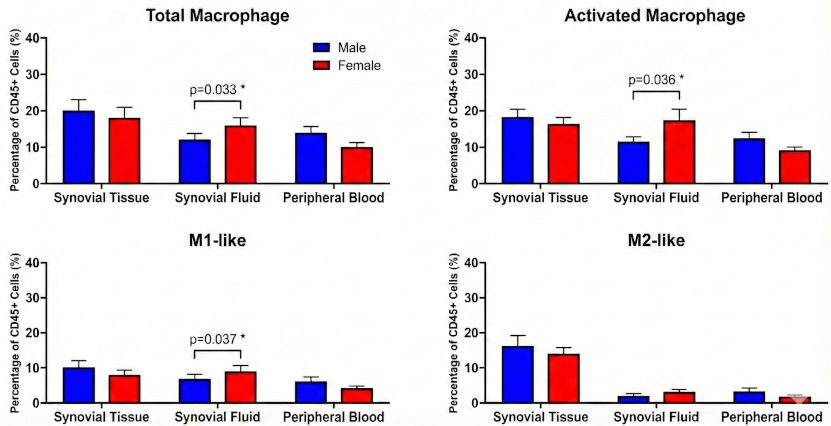
Table 1: Sex-stratified synovial fluid macrophage subpopulations in unicondylar versus bicondylar knee OA.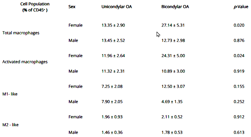
Discussion and conclusions: Sex-specific macrophage activation in late-stage knee OA was confined to the synovial fluid and emerged selectively in bicondylar disease. No sex differences were detected in unicondylar OA or in peripheral blood and synovial tissue. These findings support a sex-dependent inflammatory joint microenvironment in OA and underscore the value of sex-aware, compartment-resolved immunophenotyping for future stratification and therapeutic approaches.



