
German Congress of Orthopaedics and Traumatology 2026 (DKOU 2026)
Posterior tibial slope measurements are reliable only within a limited range of tibial rotation: A cadaveric CT-radiograph study
Text
Objectives and questions: Studies show that an elevated posterior tibial slope (PTS) leads to increased forces on the anterior cruciate ligament (ACL) causing greater anterior tibial translation and thus leading to higher failure rates following ACL reconstruction. A consensus exists for performing a slope reducing osteotomy in the setting of revision ACL reconstruction in patients with a PTS > 12°. Several factors, like length of the radiograph, knee ab-/adduction and rotation influence the lateral knee radiographs – most commonly used to evaluate the PTS. An exact correlation between tibial rotation and PTS has not yet been established. The purpose of this study was to correlate CT-Scans and radiographs of human cadaveric knees to investigate a potential correlation between tibial rotation and PTS.
Material and methods: 10 cadaveric knee joints were examined using a clinical CT-Scanner, sagittal maximum intensity projection (MIP) reconstruction were generated in the bone window perpendicular to the tibial condylar axis. Based on defined anatomical landmark–based measurements in the sagittal MIP, a neutral truelateral tibial radiographic projection was reproduced and used as the 0° reference position for subsequent radiographic examinations. Further images were then acquired in lateral projection with the specimen mounted on a rotatable device. Starting from the neutral position, radiographs were obtained with internal and external rotation of the specimen in 5° intervals up to 20°. The PTS was measured twice by an orthopedic surgeon and once by a radiologist. Measurement reliability was assessed using singlemeasure intraclass correlation coefficients for absolute agreement. Differences in PTS between observers and across rotation angles were analyzed using two-way repeated-measures ANOVA with Bonferroni post hoc correction.
Results: Intrarater reliability was excellent for the medial posterior tibial slope (ICC = 0.90) and moderate to good for the lateral slope (ICC = 0.75). Interrater reliability was good for the medial slope (ICC = 0.81) and moderate to good for the lateral slope (ICC = 0.71). For both medial and lateral PTS, measurements at 0°, 5° internal rotation, and 5° external rotation did not differ significantly. Rotations ≥10° resulted in significant systematic deviations, with medial PTS continuously decreasing by up to 4.6° in 20° external rotation and increasing by up to 3.1° in 20° internal rotation (Figure 1 [Fig. 1]), while lateral PTS changed to a lesser extent (up to −2.3° and +2.8°, respectively).
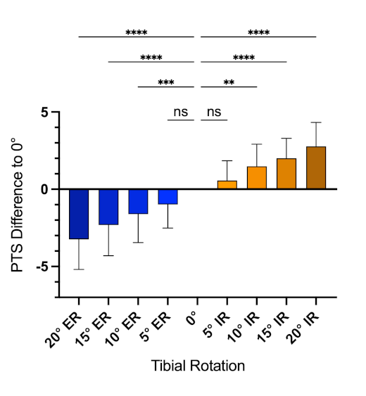
Discussion and conclusions: Posterior tibial slope measurements on lateral knee radiographs are rotation-dependent, decreasing with external rotation and increasing with internal rotation. Measurements remain stable and observer independent only within a rotational tolerance of ±5°, whereas rotations ≥10° result in systematic errors. Based on these findings, further analyses will aim to define tibia-based radiographic quality criteria for lateral knee imaging allowing reliable PTS measurement.



