German Congress of Orthopaedics and Traumatology (DKOU 2025)
Hip flexion influences cross-section-based measurement of femoral torsion: A three-dimensional comparative analysis of the Murphy and Waidelich methods
Text
Objectives and questions: Accurate assessment of femoral torsion is critical for evaluating torsional deformities and planning corrective osteotomies. It remains unclear to what extent varying degrees of hip flexion may affect torsion measurements derived from cross-sectional imaging. This experimental study aimed to determine how changes in flexion angle influence two established measurement methods (Murphy and Waidelich) and to clarify the extent to which the collum–corpus–diaphyseal (CCD) angle and baseline torsion modulate this effect.
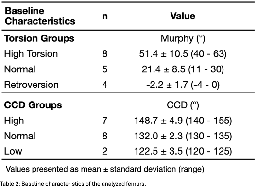
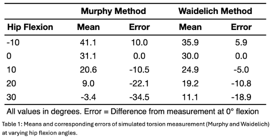
Material and methods: CT data from 17 femora were segmented and exported as three-dimensional (3D) models. Based on their torsion values (using the Murphy method on the original CT), the femora were classified as “high” (≥40°), “normal” (10–35°), or “retroversion” (≤0°). CCD angles were grouped as “high” (≥140°), “normal” (130–135°), or “low” (≤125°). In the 3D environment, femoral torsion measurements according to Murphy and Waidelich were then performed at five simulated hip flexion angles (-10°, 0°, 10°, 20°, 30°; Figure 1 A-B [Abb. 1]). Virtual axial planes were created to replicate the respective CT-based measurement techniques (Figure 1 C-D [Abb. 1]). A mixed-effects model evaluated the impact of flexion angle, measurement method, CCD group, and torsion group.

Results: Both methods showed a significant reduction in measured femoral torsion with increasing hip flexion (p<0.001). The Murphy method was almost twice as sensitive to flexion changes (mean error: -1.15° per additional degree of flexion) compared with the Waidelich method (-0.63° per degree; p<0.001). At 30° flexion, the mean torsion errors were -34.5° for the Murphy method and -18.9° for the Waidelich method (Table 1 [Tab. 1]). A high CCD angle (≥140°) further amplified this flexion-related effect, while baseline torsion did not influence it.
Discussion and conclusions: Hip flexion exerts a major influence on CT-based measurement of femoral torsion. The Murphy method is significantly more sensitive to flexion changes than the Waidelich method, and high CCD angles further increase this measurement error, while baseline torsion does not impact it. Consistent positioning of the patient with minimal hip flexion during CT acquisition is therefore crucial to maximize measurement accuracy. If this is not possible due to e.g. hip flexion contracture, multiplanar reconstruction or segmentation and subsequent 3D modelling, as introduced in this study, should be used to measure torsion orthogonal to the mechanical femur axis.
Table 2 [Tab. 2]