German Congress of Orthopaedics and Traumatology (DKOU 2025)
AI-powered 3D MRI biochemical analysis of acetabular cartilage composition in hip dysplasia and protrusion using volumetric dGEMRIC
Text
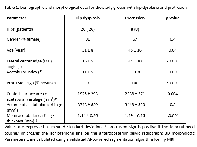
Objectives and questions: Hip dysplasia and protrusion represent opposing extremes of acetabular deformities, with dysplasia characterized by a steep acetabular inclination and reduced coverage, and protrusion by excessive coverage. These structural differences impact joint biomechanics, influencing both static loading (pressure distribution) and dynamic mechanics. Advances in medical imaging have introduced biochemical MRI techniques, such as delayed gadolinium-enhanced MRI of cartilage (dGEMRIC), which indirectly measures glycosaminoglycan (GAG) content in cartilage, with higher dGEMRIC values indicating increased GAG concentration. Artificial intelligence (AI)-powered segmentation algorithms enable automatic cartilage segmentation and incorporation of volumetric dGEMRIC evaluation (Figure 1 [Abb. 1]). Assuming that altered joint biomechanics due to different acetabular morphologies affect biochemical cartilage composition, we investigated whether the topographical distribution of dGEMRIC values differs between hips with dysplasia and protrusion.

Material and methods: This retrospective study included 34 patients (26 dysplastic hips, 8 protrusion hips) who underwent 3T MR arthrography between November 2019 and March 2023 (age and gender details in Table 1 [Tab. 1]). Imaging consisted of PD-weighted TSE and axial-oblique 3D T1 MP2RAGE MRI sequences, with anteroposterior pelvic radiographs assessed for standard parameters. Patients were classified into two groups representing extreme and opposite deformities: Protrusion (positive protrusion sign, defined as the femoral head touching or crossing the ilioischial line) and Dysplasia (LCE < 23°), based on radiographic criteria. A validated AI-powered segmentation algorithm (3D nnU-Net) was used to calculate 3D morphological parameters and dGEMRIC values per sector using a clock-face system (1–12 o’clock), further divided into central and peripheral zones. Sectors located in the area of the incisura and fossa were excluded from the analysis. Statistical analysis was performed using the Wilcoxon rank-sum test after testing for normality.
Results: Overall, dGEMRIC values did not differ between hip dysplasia and protrusion (589.9 ± 88.1 vs. 590.6 ± 152.9, p=0.9) (Table 2 [Tab. 2]). The topographical distribution was similar in both groups, with the highest values observed from 11 to 1 o’clock in both central and peripheral zones (Table 2 [Tab. 2], Figure 1 [Abb. 1]). Dysplastic hips showed greater variability in dGEMRIC values compared to protrusion hips, with both higher and lower extremes (Figure 1 [Abb. 1]).
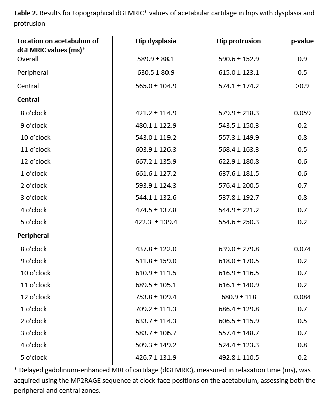
Discussion and conclusions: To our knowledge, this is the first study comparing volumetric dGEMRIC values using an AI-powered segmentation algorithm. Despite different acetabular morphologies and biomechanics in hips with dysplasia and protrusion, no topographical differences in dGEMRIC distribution were found. This suggests that biomechanical joint loading does not influence topographical biochemical cartilage composition.

