German Congress of Orthopaedics and Traumatology (DKOU 2025)
The Wnt1G177C mutation impairs fracture healing in mice
Text
Objectives and questions: Wnt1 is essential for bone development and remodeling, with pathogenic variants linked to osteogenesis imperfecta type XV and early-onset osteoporosis. However, their impact on fracture healing remains unclear. This study investigates how the Wnt1G177C/G177C variant affects inflammation, repair, and remodeling during fracture healing, potentially helping to develop targeted therapies for Wnt1-related bone disorders and osteoporosis.
Material and methods: Bone healing was assessed in 12-week-old female Wnt1+/+ and Wnt1G177C/G177C mice using a standardized femoral osteotomy stabilized with an external fixator. On day 21 post-fracture, mechanical strength was evaluated via three-point bending, and µCT measured BV/TV in the callus. Histomorphometry (day 10) analyzed tissue composition, while FACS with a 36-plex assay (day 2) quantified immune cells and chemokines/cytokines. Statistical comparisons were made using one-way ANOVA with Tukey’s test (P < 0.05, P < 0.01).
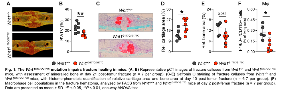
Results: To investigate the functional impact of the Wnt1G177C/G177C variant on fracture healing, a standardized femoral osteotomy was performed on 12-week-old female Wnt1+/+ and Wnt1G177C/G177C mice. After 21 days post-fracture, three-point bending testing revealed a significant reduction in mechanical strength of the left unfractured femur in Wnt1G177C/G177C mice compared to Wnt1+/+ controls (1358.38 ± 645.09 vs 439.32 ± 257.54, P < 0.01). Similar reductions were observed in the right fractured femur, with significantly lower mechanical strength in Wnt1G177C/G177C mice (50.77 ± 23.30 vs 27.08 ± 15.09, P < 0.05). µCT analysis of the unfractured femur showed a significant decrease in cortical thickness in Wnt1G177C/G177C mice compared to Wnt1+/+ controls (204.75 ± 13.11 vs 163.54 ± 9.55, P < 0.001). Similarly, µCT analysis of the fractured femur indicated a significant reduction in BV/TV in Wnt1G177C/G177C mice (Figure 1 A-B [Fig. 1]). Histomorphometric analysis of the fracture callus revealed a significant increase in relative cartilage area and a significant decrease in relative bone area in Wnt1G177C/G177C mice compared to Wnt1+/+ controls (Figure 1 C-E [Fig. 1]). Further investigation of the early inflammatory phase of fracture healing using FACS analysis demonstrated a significant decrease in macrophage numbers in Wnt1G177C/G177C mice (Figure 1 F [Fig. 1]). Cytokine and chemokine 36-plex analysis of plasma showed a significant reduction of CCL5 and CCL7 levels in Wnt1G177C/G177C mice compared to Wnt1+/+ controls.
Discussion and conclusions: The Wnt1G177C/G177C mutation impairs fracture healing by weakening mechanical strength, reducing bone formation, and disrupting early inflammatory responses. These findings underscore Wnt1’s crucial role in bone regeneration and highlight its potential as a therapeutic target for improving fracture repair in Wnt1-related disorders and osteoporosis.

