
Deutscher Rheumatologiekongress 2026
A 45-year-old female with systemic lupus erythematosus presenting with headache and fever
Text
History: A 45-year-old female presented with a 10-day history of fever (up to 38.6°C), headache, and vomiting. Her medical history included well-controlled Systemic Lupus Erythematosus (SLE) since 2019, initially characterized by photosensitive rash, peripheral arthralgias, pleuritis, Raynaud’s phenomenon, anemia, and leukopenia. Laboratory findings showed ANA (1:320), positive Anti-dsDNA, and low C3/C4 levels. The patient had been in remission for five years on low-dose corticosteroids and hydroxychloroquine.
Cardinal symptom by manifestation of disease: She was admitted to the Clinic of Internal Medicine with low-grade fever (37.7°C) and stable vitals. Neurological examination was unremarkable, with no focal signs or meningismus. Workup for infectious causes (blood/urine cultures, viral serology, syphilis, brucellosis, and tuberculosis) was negative. Subsequently, the patient developed worsening headache, blurred vision, and left temporal hemianopsia, followed by a tonic-clonic seizure managed with levetiracetam.
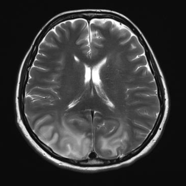
Diagnostics: Lumbar puncture revealed cerebrospinal fluid (CSF) with pleocytosis (10 cells/μL, 90%) and a protein level of 200 mg/dL. CSF cultures, FilmArray, and autoimmune/paraneoplastic panels were negative. Brain MRI demonstrated findings attributed to vasogenic edema localized in the cortical-subcortical region of the posterior structures. A diagnosis of Posterior Reversible Encephalopathy Syndrome (PRES) was established triggered by the patient’s underlying systemic autoimmune disease.
Therapy: Four biweekly (15-day) cycles of intravenous (IV) cyclophosphamide (500 mg) and one cycle of IV rituximab (two 500 mg doses, 15 days apart) were administered. Levetiracetam therapy was continued.
Further course: Two months following treatment with cyclophosphamide and rituximab, clinical improvement was observed, including improved visual acuity and visual fields on repeat ophthalmological assessments, as well as subjective visual improvement reported by the patient. Radiologically, there was regression of the vasogenic edema previously described on MRI.
Disclosures: No conflict of interest.
Figure 1 [Fig. 1]
Figure 1: Brain MRI on admission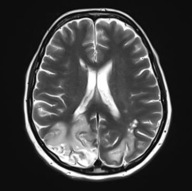
Figure 2 [Fig. 2]
Literatur
[1] Hinchey J, Chaves C, Appignani B, Breen J, Pao L, Wang A, Pessin MS, Lamy C, Mas JL, Caplan LR. A reversible posterior leukoencephalopathy syndrome. N Engl J Med. 1996 Feb 22;334(8):494-500. DOI: 10.1056/NEJM199602223340803[2] Legge AC, Hanly JG. Recent advances in the diagnosis and management of neuropsychiatric lupus. Nat Rev Rheumatol. 2024 Nov;20(11):712-728. DOI: 10.1038/s41584-024-01163-z