Deutscher Rheumatologiekongress 2025
Association of Interferon I signaling with CD4 T cell clonal expansion in rheumatoid arthritis
Text
Introduction: The T cell repertoire of patients with rheumatoid arthritis (RA) is characterized by the expansion of large clones, possibly developed due to chronic (auto-)antigen stimulation [1]. A comprehensive study of clonally expanded T cells could shed light on mechanisms of T-cell-mediated autoimmunity in RA.
Methods: Single-cell sequencing of RNA, T cell receptor (TCR) and hashtags for CD4 and CD8 surface expression was performed in circulating T cells (RA n=3; healthy donors, HD n=2). Circulating T cells were isolated from blood samples for flow cytometry analysis (RA n=14, HD n=12). For 4 additional RA patients, blood and synovial fluid samples were available.
Results: Clonality analysis revealed that in contrast to healthy individuals, RA patients have large T cell clones (³10 T cells with the same paired TCR sequence) more frequently in the CD4 than in the CD8 compartment (CD4 in 92.3% vs. 23.5% of all large T cell clones in RA vs. HD; p < 0.001). Interestingly, lack of CCR7 and CD27 expression is found in 94% and 85% of large clones in RA and HD, respectively, making this combination a useful global marker for large clonal expansion.
We identified 1,651 genes differentially expressed in RA vs. HD clonally expanded CD4 T cells. Gene ontology analysis revealed type I interferon pathway (JAK1, IFITM2, STAT4) and granzyme-mediated signaling (GZMB, PRF1, FGFBP2) to be upregulated, whereas HLA-class-II expression (HLA-DRB1, HLA-DRB5, HLA-DRA) was downregulated in RA vs. HD expanded CD4 T cells (Figure 1 [Fig. 1]).
Figure 1: Differentially expressed genes in clonally expanded CD4 T cells of RA patients vs. healthy controls.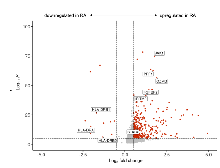
Flow cytometry analysis showed that CCR7-CD27- T cells accumulate in the joints of RA patients compared to the peripheral blood (29.5% vs. 5.5% of total T cells; p = 0.03). Validation of selected markers using flow cytometry confirmed Perforin to be significantly upregulated in RA compared to HD CCR7-CD27- T cells (37.9% vs 5.2% Perforin+ T cells; p = 0.001).
Conclusion: Expansion of large T cell clones in RA predominantly occurs in the CD4 T cell compartment. Clonally expanded CD4 T cells in RA accumulate in the joint, upregulate interferon-related genes and are thus likely to contribute to inflammation.

