German Congress of Orthopaedics and Traumatology (DKOU 2025)
A new era of hip fracture surgical outcomes: AI-driven individual muscle segmentation and functional recovery prediction
Text
Objectives and questions: Total hip arthroplasty (THA) is a widely used surgical intervention for hip fractures, yet post-surgical recovery remains highly variable. Assessing muscle volume (MV) and intramuscular adipose tissue (IMAT) is essential for understanding rehabilitation outcomes. However, manual segmentation of individual muscles from CT scans is time-consuming and subject to variability. This study aims to develop an AI-based automatic segmentation model to quantify MV and IMAT changes following THA. By analyzing individual thigh muscles preoperatively and at multiple follow-up points, this study seeks to determine which specific muscles experience the greatest volume loss and fat infiltration. Additionally, the study investigates whether bilateral asymmetry in muscle volume and IMAT correlates with post-surgical functional recovery.
Material and methods: This study included 50 patients aged 50 years or older who underwent THA. Whole-thigh CT scans (L3 to the knee joint) were analyzed using a UNETR-based AI model (Dice score = 0.91). Segmentation targeted individual muscles, including (Figure 1 [Abb. 1]):
Figure 1: Pipeline of individual thigh muscle status evaluation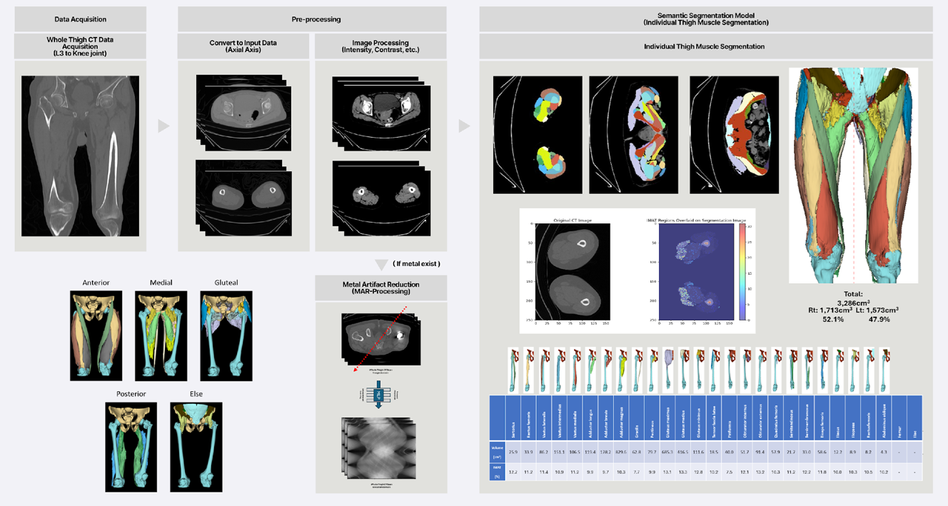
Anterior compartment: Vastus lateralis, vastus intermedius, vastus medialis, rectus femoris, sartorius.
Medial compartment: Adductor longus, adductor brevis, adductor magnus, gracilis.
Posterior compartment: Semitendinosus, semimembranosus, biceps femoris.
Hip region: Gluteus maximus, gluteus medius, gluteus minimus.
MV and IMAT percentage were quantified bilaterally at preoperative, postoperative, 3-month, 6-month, and 1-year follow-ups. A subset of participants underwent gait analysis using smart insoles, OpenPose-based motion tracking, and electromyography (EMG) sensors. Differences in MV and IMAT between the operated and non-operated limbs were analyzed to assess their correlation with post-surgical recovery.
Results: Significant muscle volume reduction was observed on the fracture side, particularly in the vastus lateralis, vastus intermedius, vastus medialis, rectus femoris, adductor longus, and gluteus maximus. Increased IMAT was strongly correlated with muscle atrophy across all thigh muscle compartments (p < 0.01). In contrast, most hip muscles, except gluteus maximus, exhibited no significant IMAT changes. Patients with greater side-to-side muscle imbalance showed poorer functional recovery.
Discussion and conclusions: AI-driven automatic muscle segmentation enables precise, longitudinal tracking of muscle volume loss and fat infiltration following THA. This approach provides a data-driven framework for surgical planning and rehabilitation, offering insights into side-by-side asymmetry and functional recovery trajectories. Understanding muscle-specific degeneration patterns facilitates personalized rehabilitation strategies, improving post-surgical outcomes for hip fracture patients. Future studies integrating gait biomechanics and long-term follow-up are needed to optimize post-THA rehabilitation interventions.

