
Deutscher Rheumatologiekongress 2026
Targeting the vascular adhesion protein-1/Siglec-9 axis in giant cell arteritis
Text
Introduction: The precise pathogenic mechanisms facilitating localized vascular inflammation in giant cell arteritis (GCA) remains incompletely understood. Vascular Adhesion Protein-1 (VAP-1) and its leukocyte ligand, Sialic Acid Binding Ig-like Lectin 9 (Siglec-9), govern critical steps in leukocyte extravasation. This study aimed to characterize the VAP-1/Siglec-9 axis in GCA and evaluate the diagnostic feasibility of [68Ga] Ga-DOTA-Siglec-9 positron emission tomography computed tomography (PET/CT) for detecting active vasculitis.
Methods: A prospective cohort of patients with GCA and age- and sex-matched healthy control subjects was recruited at the Division of Rheumatology, University Hospital Bonn. Tissue expression of VAP-1 was assessed via immunohistochemistry in temporal artery biopsies (TABs). Soluble VAP-1 (sVAP-1) and Matrix Metalloproteinases (MMPs) were quantified in serum via Enzyme-linked Immunosorbent Assay. Peripheral blood mononuclear cells (PBMCs) were analyzed utilizing flow cytometry to determine Siglec-9 surface expression and bulk RNA-sequencing to define transcriptomic signatures. Molecular imaging was performed in a subset of active GCA patients using [68Ga] Ga-DOTA-Siglec-9 PET/CT, with follow-up scans acquired two hours post-glucocorticoid administration. Furthermore, a cohort of GCA patients in stable remission was subjected to transcriptomic profiling.
Results: Histopathological analysis revealed marked VAP-1 expression localized (in addition to the staining reaction in the vascular smooth muscle cells) specifically to the hyperplastic neointima of inflamed GCA arteries (Figure 1A-C [Fig. 1]). Conversely, circulating sVAP-1 (Figure 1D [Fig. 1]) and its shedding enzymes MMP-2 (Figure 1E [Fig. 1]) and MMP-9 (Figure 1G [Fig. 1]) were decreased, whereas pro-inflammatory MMP-3 (Figure 1F [Fig. 1]) was elevated in active GCA patients. Flow cytometry demonstrated significant upregulation of Siglec-9 on classical and non-classical monocytes, plasmablasts, plasma cells, and natural killer cells (Figure 1H [Fig. 1]). Bulk RNA-sequencing revealed a profound proinflammatory signature, extensive myeloid skewing, and a deficit in regulatory T cells (Figure 1J [Fig. 1]). An in vivo, [68Ga] Ga-DOTA-Siglec-9 PET/CT exhibited a localized tracer uptake in the thoracic and abdominal aorta and axillary arteries (not shown). Transcriptomic analysis of patients in stable remission identified a distinct molecular signature driven by dysregulated cell adhesion and wound-healing networks, rather than a return to homeostatic baseline.
Figure 1: Dysregulation of the VAP-1/Siglec-9 Axis and systemic immune remodeling in active giant cell arteritis. (A–C) Representative micrographs of temporal artery biopsies. (A) Hematoxylin and Eosin (H&E) staining of active GCA. (B, C) Immunohistochemical staining for VAP-1 in an inflamed artery demonstrating intense expression. (D) Soluble VAP-1 (sVAP-1), (E) Matrix metalloproteinase-2 (MMP-2), (F) Matrix metalloproteinase-3 (MMP-3), and (G) Matrix metalloproteinase-9 (MMP-9) concentrations (1). (H) Flow cytometric quantification of Siglec-9 positive cells (%) across distinct PBMCs subsets in active disease (orange) compared to controls (gray). (J) Stacked bar plots illustrating deconvolutions of relative frequencies of circulating immune cell populations in newly diagnosed patients compared to controls.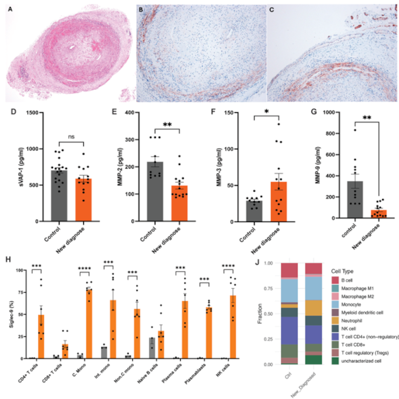
Conclusion: The VAP-1/Siglec-9 axis is highly dysregulated in active GCA, bridging localized endothelial activation with systemic myeloid expansion. [68Ga] Ga-DOTA-Siglec-9 PET/CT serves as a sensitive, dynamically responsive molecular imaging modality for VAP-1 mediated vascular inflammation. Furthermore, stable disease remission is characterized by persistent transcriptional alterations in cell adhesion pathways, highlighting an incomplete restoration of immune homeostasis.