
Deutscher Rheumatologiekongress 2026
Robot-assisted arthrosonography (ARTHUR) with AI analysis (DIANA) for the initial diagnosis and follow-up of rheumatoid arthritis: Real-world data from 300 patients with suspected arthritis
Text
Introduction: Epidemiological data indicate an increased prevalence of inflammatory rheumatic diseases (IRD) [1]. For example, the global incidence of rheumatoid arthritis (RA) has risen from 11.66 in 1990 to 13.48 per 100,000 people in 2022 [2]. Early diagnosis and initiation of therapy are essential to prevent permanent structural damage and comorbidities in RA patients [3]. However, in some states, such as Schleswig-Holstein (SH) in northern Germany, there are differences in the availability of rheumatological care between urban and rural areas. Consequently, new models of early consultation and diagnostic and therapeutic approaches using AI-trained systems are needed. Automated robotic arthrosonography has demonstrated promising potential for the detection of IRD. Nevertheless, existing evidence is limited by the small number of studies conducted to date [3].
This study, which was initiated by the patient organisation Rheuma-Liga SH, aimed to evaluate the real-world use of fully automated robotic arthrosonography (ARTHUR) with AI analysis (DIANA) to detect arthritis activity.
Methods: Between October 2024 and November 2025, we set up a mobile contact point in various communities in SH. Using ARTHUR, we detected arthrosonographic signs of new-onset or relapsing arthritis according to the OMERACT-EULAR synovitis criteria [4]. Data were collected from participants with suspected new-onset or relapsing Arthritis regarding their age, sex, morning stiffness, tender joint count (TJC), swollen joint count (SJC), and pain level on a numeric pain scale. CRP were measured using an AFIAS POC system. Health Assessment Questionnaire (HAQ-II) and Disease Activity Score-28 (DAS28) were also assessed. Participants with positive results, according to the DIANA AI analysis, were referred directly to a rheumatology specialist. Furthermore, participants with elevated CRP and/or severe symptoms were also referred to a rheumatology specialist after automated arthrosonography, even if the result was negative.
Results: A total of 420 participants underwent ARTHUR screening. Of these, 119 were excluded due to missing data. One measurement failed. The remaining 300 participants were divided into two groups: those without a history of IRD (non-IRD; n=255; 1.912 joints; age: 62.0±12.2 years; 79.1% female; DAS28:3.7±1.1; HAQII:1.1±1.1; CRP:7.1±8.5 mg/L) and those with IRD (RA: n=40; 300 joints; PMR: n=5; SSc: n=1; SLE: n=1; GPA: n=1; psoriatic arthritis: n=3; age: 65.2±13.5 years; 86.7% female; DAS28:3.6±1.3; HAQ II:1.4±1.6; CRP: 7.2±6.4 mg/L). In the non-IRD group, 41 individuals tested positive for signs of synovitis. According to a rheumatologist, five of these participants had false positive results. Only one person with a negative ARTHUR result had received a false negative result (ROC-AUC: 0.9977, sensitivity 98.99%, specificity 100%, P<0.0001, number needed to diagnose:1.01) (Figure 1 [Fig. 1]). Only patients with a suspected RA relapse were further investigated in the IRD group. Here, 6 RA patients with active disease were detected by ARTHUR, without any false positive results. Furthermore, in this group, there were no false negative results identified by a rheumatologist (ROC-AUC: 1.000, sensitivity 100%, specificity 100%, P=0.0016, number needed to diagnose: 1.010) (Figure 1 [Fig. 1]).
Figure 1: Sensitivity and specificity of ARTHUR in identifying arthritis in participants without a history of IRD (non-IRD, left) and in RA patients suspected of relapse (right).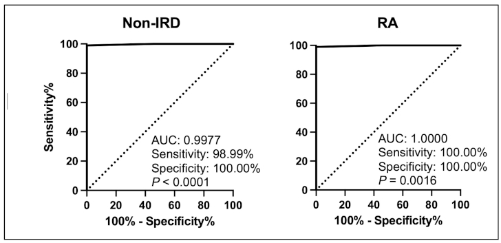
Conclusion: Fully automated robot-assisted arthrosonography with AI analysis demonstrated high sensitivity and specificity in detecting arthritis in patients with new-onset disease as well as in RA patients suspected of relapse.
Disclosures: The authors declare no competing interests.
Literatur
[1] Bournia VK, Fragoulis GE, Mitrou P, Tsolakidis A, Mathioudakis K, Vassilopoulos D, Tektonidou M, Paraskevis D, Sfikakis PP. Increased prevalence of inflammatory arthritis, systemic lupus erythematosus and systemic sclerosis, during 2020-2023 versus 2016-2019 in a Nation-Wide Cohort Study. Rheumatol Int. 2024 Dec;44(12):2837-2846. DOI: 10.1007/s00296-024-05733-y[2] Zhang Z, Gao X, Liu S, Wang Q, Wang Y, Hou S, Wang J, Zhang Y. Global, regional, and national epidemiology of rheumatoid arthritis among people aged 20-54 years from 1990 to 2021. Sci Rep. 2025 Mar 28;15(1):10736. DOI: 10.1038/s41598-025-92150-1
[3] Frederiksen BA, Hammer HB, Terslev L, Ammitzbøll-Danielsen M, Savarimuthu TR, Weber ABH, Just SA. Automated ultrasound system ARTHUR V.2.0 with AI analysis DIANA V.2.0 matches expert rheumatologist in hand joint assessment of rheumatoid arthritis patients. RMD Open. 2025 Aug 5;11(3):e005805. DOI: 10.1136/rmdopen-2025-005805
[4] D'Agostino MA, Terslev L, Aegerter P, Backhaus M, Balint P, Bruyn GA, Filippucci E, Grassi W, Iagnocco A, Jousse-Joulin S, Kane D, Naredo E, Schmidt W, Szkudlarek M, Conaghan PG, Wakefield RJ. Scoring ultrasound synovitis in rheumatoid arthritis: a EULAR-OMERACT ultrasound taskforce-Part 1: definition and development of a standardised, consensus-based scoring system. RMD Open. 2017 Jul 11;3(1):e000428. DOI: 10.1136/rmdopen-2016-000428