Deutscher Rheumatologiekongress 2025
Gene expression analysis reveals distinct immune pathways in the synovial membrane of male RA patients versus healthy males
Text
Introduction: Rheumatoid arthritis (RA) is a complex autoimmune disease with significant immune and metabolic dysregulation [1], [2]. While most research has focused on female patients, male RA patients exhibit unique immune characteristics that remain poorly understood. This study aims to investigate the immune and metabolic features of male RA patients by analyzing mitochondrial protein expression in T and B cells and performing transcriptomic analysis to compare RA male patients with healthy male controls.
Methods: We utilized existing total RNA transcriptome data from the GEO database (GSE89408), performed on joint synovial biopsies from subjects with and without RA. The study involved the identification of differentially expressed genes (DEGs) using bioinformatics tools, followed by KEGG and GO enrichment analysis. Ex vivo protein expression levels of TOMM20 and MT-ND1 in T cells and B cells from male patients were determined by Western blot (WB) analysis.
Results: WB analysis showed no significant difference in TOMM20 expression between T and B cells. MT-ND1 expression was slightly higher in B cells compared to T cells, but the difference was not statistically significant, suggesting that both T and B cells might have similar mitochondrial metabolism in RA, as shown in Figure 1 [Fig. 1]. Bioinformatics analysis identified differentially expressed genes (DEGs) in male RA patients, with GO enrichment analysis highlighting pathways such as cytokine activity, immune receptor activity, chemokine binding, antigen binding, and complement activation. KEGG analysis revealed significant involvement in immune-related pathways, including B cell receptor signaling and phagocytosis, indicating altered immune regulation in the synovial membrane of male RA patients, as shown in Figure 2 [Fig. 2].
Figure 1: Western blot analysis of mitochondrial proteins in T and B cells from male RA patients and healthy male controls. (A) WB bands showing the expression of TOMM20 and MT-ND1 in isolated T and B cells. HSP90 was served as a loading control. (B) Quantification of protein expression levels relative to HSP90. No significant difference was observed in TOMM20 expression between T and B cells. Data are presented as mean ± standard deviation (SD). Analysis was performed using Student t-test (p < 0.05 was considered significant).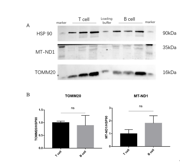
Figure 2: GO enrichment analysis of differentially expressed genes in the synovial membrane of male RA Patients when compared with healthy (non-arthritis) donors.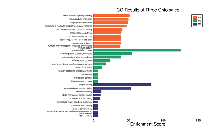
Conclusion: These findings suggest that regardless of their distinct immune functions both T and B cells in male RA patients have comparable mitochondrial metabolism. Whether this differs from healthy male individuals and female RA patients must be further explored. Nonetheless, the transcriptomic analysis of the synovial membrane clearly shows an immune dysregulation in RA, particularly in cytokine signaling and B cell activation. This study provides new insights into the immune-metabolic landscape of male RA patients and highlights potential pathways for further investigation in RA pathogenesis and targeted therapy.
Literatur
[1] Weyand CM, Goronzy JJ. Immunometabolism in early and late stages of rheumatoid arthritis. Nat Rev Rheumatol. 2017 May;13(5):291-301. DOI: 10.1038/nrrheum.2017.49[2] Gravallese EM, Firestein GS, Koscal N, Ling E, Longo DL, Messenger LA, Schubach A. What Is Rheumatoid Arthritis? N Engl J Med. 2024 Apr 4;390(13):e32. DOI: 10.1056/NEJMp2310178

