
Deutscher Kongress für Orthopädie und Unfallchirurgie 2026 (DKOU 2026)
Bioactive calcium sulfate/hydroxyapatite augmentation with systemic zoledronic acid for pedicle screw fixation in an osteoporotic minipig model
Text
Objectives and questions: Pedicle screw fixation in osteoporotic bone remains biomechanically challenging (Tian et al., 2022). While PMMA cement provides immediate stability, it is bioinert and does not promote osseointegration (Tian et al., 2022). Calcium sulfate/hydroxyapatite (CaS/HA) composites represent bioactive alternatives (Tian et al., 2022), [1], and systemic bisphosphonates such as zoledronic acid (ZA) may further enhance peri-implant bone formation [2]. This study investigated whether systemic ZA enhances the fixation strength of pedicle screws augmented with CaS/HA in an osteoporotic large-animal model.
Material and methods: A controlled prospective in vivo animal study was conducted using eleven female Göttingen minipigs. Osteoporosis was induced via ovariectomy combined with glucocorticoid administration and a calcium-restricted diet. After 6 months, pedicle screws (4.5 × 25 mm) were inserted into five lumbar vertebrae. One side was augmented with approximately 0.5 mL CaS/HA cement, while contralateral pedicles served as non-augmented controls. Systemic ZA was administered one-week post-implantation. Specimens were harvested at 2, 6, and 12 weeks (n=3/timepoint). µCT and synchrotron CT assessed peri-implant bone structure. Bone volume fraction (BV/TV) and bone mineral density (BMD) were quantified in defined ROIs. Biomechanical fixation was evaluated by manually measured maximum extraction torque. Histology (H&E, Goldner) assessed tissue response. Statistical analysis included Shapiro-Wilk testing and paired t-tests (p<0.05).
Results: µCT confirmed successful osteoporosis induction, with significantly reduced BV/TV and BMD in OVX animals compared with sham controls after 6 months (Figure 1A [Fig. 1]). Synchrotron CT following screw removal demonstrated peri-thread bone formation. Residual bone substitute was present at the screw tips at 2 and 6 weeks but was largely resorbed by 12 weeks (Figure 1B [Fig. 1]). Quantitative µCT revealed significantly higher peri-implant BV/TV on the augmented side compared with screw-only controls (p<0.05) (Figure 1C [Fig. 1]). No differences were observed in manually measured maximum removal torque between groups (Figure 1D [Fig. 1]). Histology confirmed the presence of biomaterial remnants at 6 weeks, whereas at 12 weeks, the CaS/HA was largely resorbed, with no additional new bone formation (Figure 1E [Fig. 1]).
Figure 1: Imaging, biomechanical, and histological evaluation of pedicle screw augmentation.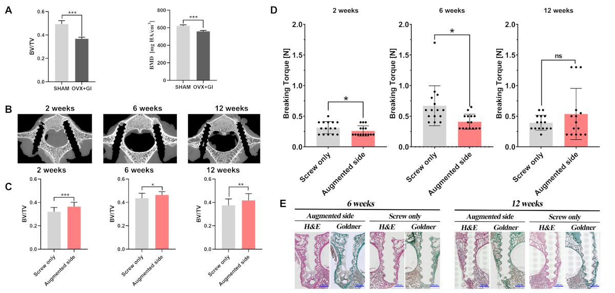
Discussion and conclusions: Systemic ZA combined with CaS/HA augmentation increased peri-implant bone volume but did not improve biomechanical fixation in this osteoporotic minipig model. Limited bioactivation in cortical-dominant pedicles and anatomical constraints may explain the absent mechanical benefit. Further studies in models more closely reflecting human spinal anatomy and loading are required to validate the translational potential of bioactive augmentation strategies.
Literatur
[1] Tian X, et al. Comparison of immediate anchoring effectiveness of two different techniques of bioresorbable ceramic application for pedicle screw augmentation. Ceramics International. 2024;50(8):12877-12889. DOI: 10.1016/j.ceramint.2024.01.195[2] Markeviciute V, Puthia M, Arvidsson L, Liu Y, Törnquist E, Tengattini A, Huang J, Bai Y, Vater C, Petrolis R, Zwingenberger S, Krisciukaitis A, Smailys A, Lukosevicius S, Stravinskas M, Isaksson H, Tarasevicius S, Lidgren L, Tägil M, Raina DB. Systemically administered zoledronic acid activates locally implanted synthetic hydroxyapatite particles enhancing peri-implant bone formation: A regenerative medicine approach to improve fracture fixation. Acta Biomater. 2024 Apr 15;179:354-370. DOI: 10.1016/j.actbio.2024.03.005



