
Deutscher Kongress für Orthopädie und Unfallchirurgie 2026 (DKOU 2026)
Bone healing by tissue-loaded scaffolds using BoneFlo® in a critical size defect ovine model
Text
Objectives and questions: Surgical reconstruction of critical‑size defects (CSD), remains one of the most demanding procedures in orthopaedics. Besides autologous bone grafting, synthetic graft material is one option to address these defects. BoneFlo® is a system which enables the entrapment of autologous tissue in a porous synthetic bone scaffold during the operating procedure, representing a promising approach to improve the bone forming potential in CSD. Hence, the aim of this study was to (1) assess the healing of an established critical-size tibial defect in an ovine model [1] treated with 3D-printed medical-grade polycaprolactone/β-tricalcium phosphate (mPCL-TCP) scaffold loaded with autologous tissue harvested with an innovative suction handle & to (2) compare it to the healing behavior with pristine mPCL-TCP scaffolds.
Material and methods: A total of 16 sheep were divided into two groups: 14 animals received a vacuum filtered scaffold by autologous tissue released at the surgical site during tibial osteotomy (VFS), whereas 2 sheep received pristine scaffolds (control group; PS). A tubular porous bone scaffold made of mPCL-TCP (Osteopore, Singapore) was used in all cases. For VFS the scaffold was placed as an internal filter in a BoneFlo® (TissueFlow, Germany) suction device intra-operatively & loaded with autologous tissue by suctioning during tibia osteotomy. Moreover, small fragments from the resected bone were added harvested by a bone mill. A standard 3 cm defect was set in the tibia, filled with either of the scaffolds & additionally stabilized with a plate. Bone healing was assessed blinded using X-rays at 2 weeks, 3 & 6 months post-operatively. In addition, this study was accomplished by human in vitro data.
Results: All operated sheep had an uneventful recovery from general anesthesia and surgery. In contrast to pristine scaffolds (PS) we found significant increased new bone formation in all tissue activated mPCL-TCP groups (VFS) within 3 months. In 13 of 14 animals (93%) the CSD healed completely within 6 months. Interestingly, bone regeneration was identified not only at the adjacent diaphyseal osteotomy sites, but also within the scaffolds (Figure 1 [Fig. 1]). The CSD size defect was overbridged by bony tissue involving the cortical bone. In contrast, we found no bone healing & only small amounts of bone formation in untreated controls (PS). Moreover, cell cultural experiments confirmed the high regenerative nature of surgical site released tissue cultivated onto mPCL-TCP (proliferation, osteoblastic differentiation).
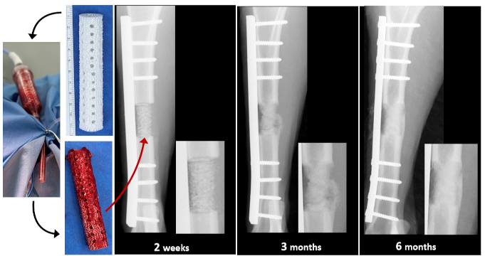
Discussion and conclusions: The results of this study showed significant bone formation within a 3D-fabricated mPCL-TCP scaffold in a critical size tibial ovine defect after impregnation with autologous surgical site released tissue. These data were supported by our in vitro results and correspond to the findings of other investigators [2], [3], underlining the great osteogenic nature of this composite.
Literatur
[1] Sparks DS, Saifzadeh S, Savi FM, Dlaska CE, Berner A, Henkel J, Reichert JC, Wullschleger M, Ren J, Cipitria A, McGovern JA, Steck R, Wagels M, Woodruff MA, Schuetz MA, Hutmacher DW. A preclinical large-animal model for the assessment of critical-size load-bearing bone defect reconstruction. Nat Protoc. 2020 Mar;15(3):877-924. DOI: 10.1038/s41596-019-0271-2[2] Rehage E, Sowislok A, Busch A, Papaeleftheriou E, Jansen M, Jäger M. Surgical Site-Released Tissue Is Potent to Generate Bone onto TCP and PCL-TCP Scaffolds In Vitro. Int J Mol Sci. 2023 Nov 1;24(21):15877. DOI: 10.3390/ijms242115877
[3] Groven RVM, Blokhuis JT, Poeze M, van Griensven M, Blokhuis TJ. Surgical suction filter-derived bone graft displays osteogenic miRNA and mRNA patterns. Eur J Trauma Emerg Surg. 2024 Feb;50(1):315-326. DOI: 10.1007/s00068-023-02350-5



