Deutscher Kongress für Orthopädie und Unfallchirurgie 2026 (DKOU 2026)
Functional knee stability across the menstrual cycle: A prospective investigation of dynamic landing biomechanics
Text
Objectives and questions: Anterior cruciate ligament (ACL) ruptures occur two to eight times more frequently in physically active women than in men [1]. This increased risk is considered multifactorial, involving anatomical, neuromuscular and biomechanical factors, with a potential contribution of hormonal fluctuations. While menstrual cycle–dependent changes in knee ligament laxity have been reported [2], ligament laxity alone does not seem to sufficiently explain the observed injury incidence [3]. Yet, a peak in ACL injury incidence has been registered around ovulation [4], suggesting that factors other than tissue composition might be affected by estrogen-level changes. The purpose of this study is to investigate menstrual cycle–related differences in dynamic knee joint stability using objective biomechanical parameters during a functional landing task.
Material and methods: In this prospective observational study, eumenorrheic, physically active women without a history of knee injury are included. Biomechanical tests are performed at two points within a menstrual cycle: during the early follicular phase (days 2–4) and around ovulation. Ovulation is determined using cycle tracking combined with luteinizing hormone urine tests, as recommended in previous biomechanical studies [5]. Participants perform standardized single-leg drop jump tasks onto force plates. Biomechanical analysis is performed using a 3D motion capture system for marker-based kinematic and kinetic evaluation. Primary outcome measures are peak vertical ground reaction force (peak vGRF) and frontal plane knee valgus angle during landing, both of which are established surrogates of ACL injury risk [6]. Secondary outcomes include time to stabilization of ground reaction forces and ground contact time. Outcomes are analyzed intraindividually between cycle phases using paired statistical tests (p < 0.05).
Results: Phase-dependent differences in peak vGRF and frontal plane knee valgus angle between the early follicular and ovulatory phase are currently being analyzed, based on prior evidence of cycle-related alterations in landing biomechanics [5,6]. Time to stabilization and interindividual variability in the magnitude of biomechanical changes across cycle phases are likewise being evaluated [7].
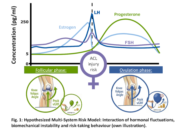
Discussion and conclusions: This study addresses the need for objective biomechanical markers of cycle-dependent ACL injury risk. If confirmed, significantly elevated peak vGRF and knee valgus angles during ovulation would suggest that ACL injury risk is driven by transient compromises in dynamic neuromuscular control rather than passive laxity alone, consistent with multifactorial injury models described in the literature [1]. Such findings would challenge the isolated focus on anatomical factors. Future studies should explore whether biomechanical instability and cycle-related changes in risk-taking behaviour interact within a multi-system model of ACL injury risk (Figure 1 [Fig. 1]).