Deutscher Rheumatologiekongress 2026
Regulation of endothelial vascular adhesion protein-1 in giant cell arteritis
Text
Introduction: Vascular adhesion protein-1 (VAP-1) is an endothelial cell adhesion molecule possessing enzymatic activity, functioning critically in leukocyte extravasation and vascular inflammation. Its pathophysiological significance in systemic vasculitis including giant cell arteritis (GCA) remains insufficiently characterized. This study aimed to investigate endothelial VAP-1 expression in human aortic endothelial cells (HAoEC), and the immunomodulatory influence of glucocorticoid therapy.
Methods: Human aortic endothelial cells (HAoEC) and human umbilical vein endothelial cells (HUVEC, CRL-1730, ATCC) were cultured and stimulated with proinflammatory mediators, including Tumor Necrosis Factor-alpha (TNF-a), Interferon-gamma (IFN-g), and Lipopolysaccharides (LPS), at varying concentrations i.e. 10, 100 µg/mL and time intervals of 24 and 48 hours. Subsequently, VAP-1 surface expression was measured via flow cytometry. The immunomodulatory effect of prednisolone on cytokine induced VAP-1 expression was evaluated in parallel stimulation setups and statistically analyzed.
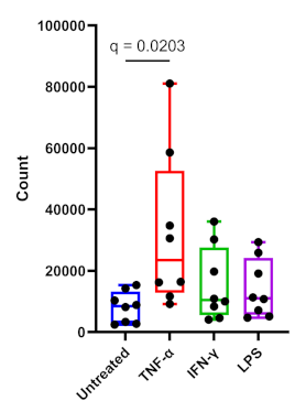
Results: Stimulation of HAoEC cells with proinflammatory mediators induced pronounced VAP-1 expression. TNF-a exhibited the most potent effect, resulting in approximately 80 to 90% VAP-1+ cells at a concentration of 100 µg/mL (p ≤ 0.02). IFN-g and LPS also induced significant upregulation of VAP-1 expression, albeit to a lesser extent than TNF-a. Dose response relationships were observed for all stimuli. Conversely, prednisolone treatment markedly attenuated cytokine induced VAP-1 expression on endothelial cells across all stimulation conditions. Comparison between HAoEC and HUVEC revealed no significant differences in VAP-1 expression under basal or stimulated conditions.
Conclusion: This study demonstrated that endothelial VAP-1 expression is induced by proinflammatory stimuli and effectively suppressed by glucocorticoid therapy. Additionally, VAP-1 is significantly upregulated in temporal arteries of patients with GCA. These findings support a functional role for VAP-1 in vascular inflammatory responses and underscore its potential utility as a biomarker and therapeutic target in systemic vasculitis such as GCA.
Figure 1 [Fig. 1]
Figure 1: The frequency of VAP-1-positive cells under diverse inflammatory conditions is depicted. Stimulation with Tumor Necrosis Factor-alpha (TNF-a) (p = 0.02), Interferon-gamma (IFN-?), and Lipopolysaccharides (LPS) induced a significant upregulation of expression relative to the unstimulated control.