
Deutscher Kongress für Orthopädie und Unfallchirurgie 2026 (DKOU 2026)
Integrated in vivo and in vitro transcriptomics reveal inflammatory drivers of GC-induced bone loss
Text
Objectives and questions: Glucocorticoids (GCs) are widely used anti-inflammatory drugs; however, high-dose or long-term administration is a major cause of secondary osteoporosis, known as glucocorticoid-induced osteoporosis (GIO). Despite its clinical relevance, the molecular mechanisms by which GCs suppress osteoblast differentiation and impair bone formation are not fully understood. To address this gap, we employed an integrated in vivoandin vitrotranscriptomic approach to identify shared molecular drivers of GC-induced bone loss.
Material and methods: Wild-type BALB/c mice received subcutaneous implantation of slow-release vehicle- or prednisolone-pellets (12 mg/kg/day) for two weeks to induce GIO. High-resolution micro-computed tomography (µCT) was performed on explanted femora to assess trabecular and cortical bone parameters. Efficient induction of GIO was further verified by histomorphometric analysis of bone sections and plasma ELISAs. Primary murine osteoblasts were isolated from bone marrow-depleted long bones using manual chopping and enzymatic digestion. Cells were cultured in osteogenic induction medium (OI) or OI supplemented with 1 µM dexamethasone (DEX). On day 6 post-induction, gene expression was analyzed by qRT-PCR, and alkaline phosphatase (Alp) staining was used to assess osteoblast differentiation. RNA sequencing (RNAseq) was performed on RNA isolated from bone marrow-cleared humeri and from cultured primary murine osteoblasts. Statistical analyses were conducted using an unpaired Student’s t-test.
Results: In vivo prednisolone treatment markedly reduced trabecular and cortical bone mass (Figure 1A [Fig. 1]). This bone loss was associated with impaired osteoblastogenesis (Ob.N/B.Pm: 5.02±1.10 vs 0.99±0.58) and enhanced osteoclastogenesis (Oc.N/B.Pm: 8.08±1.98 vs 11.93±2.94), consistent with decreased PINP levels (4.89±1.02 vs 2.48±0.67 ng/mL) and increased CTX-I levels (34.52±5.94 vs 49.01±7.44 ng/mL). In parallel, DEX-treated primary osteoblasts exhibited reduced Alp activity (Figure 1B [Fig. 1]) and downregulation of osteoblast marker genes (Sp7, Alpl). Integrated analysis ofin vivoandin vitroRNAseq datasets (Figure 1C [Fig. 1]), followed by Metascape enrichment analysis, revealed strong enrichment of inflammatory response-related pathway (Figure 1D [Fig. 1]). Within this pathway, Saa3 emerged as one of the most highly upregulated genes.The increased expression of Saa3 induced by high-dose GCs suggests that the detrimental effects of GCs observed bothin vitroandin vivomay be mediated, at least in part, through Saa3 upregulation, which needs further investigation.
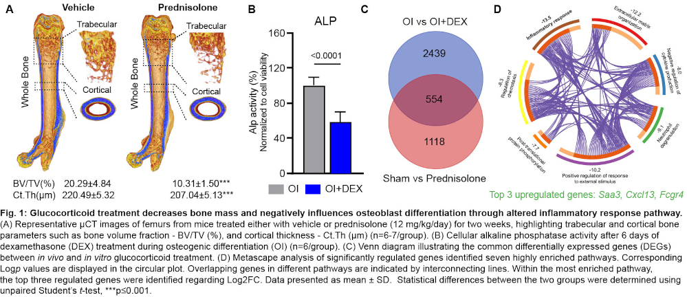
Discussion and conclusions: Integrated transcriptomic analyses revealed activation of inflammatory pathways, with Saa3 emerging as a potential key mediator. GC-induced Saa3 expression may inhibit osteoblast differentiation, thereby linking inflammatory signaling to suppressed bone formation. These findings highlight Saa3 as a potential therapeutic target to mitigate bone loss and reduce fracture risk, although further investigation is required to establish its mechanistic role.



