
Deutscher Kongress für Orthopädie und Unfallchirurgie 2026 (DKOU 2026)
In vitro cellular and histopathological effects of patch preparation techniques on human long head of the biceps tendon tissue – preliminary results
Text
Objectives and questions: High retear rates after arthroscopic rotator cuff repair are partly attributed to compromised tendon biology. Autologous augmentation using the long head of the biceps tendon (LHBT) has emerged as a biologically active scaffold; however, LHBT-based patch techniques require mechanical manipulation that may affect cellular viability and tissue integrity. This study evaluates the in vitro cellular, molecular, and histopathological effects of different long head of the biceps tendon (LHBT) autograft preparation techniques.
Material and methods: This prospective single-center tissue analysis study analyzes LHBT tissue harvested during routine shoulder surgery in patients undergoing planned biceps tenotomy and tenodesis without intraoperative signs of macroscopic tendinopathy. Tendon samples from up to 30 patients are divided into three preparation conditions: native (untreated) tendon, sliced tendon patch, and pressed tendon patch. Cellular activity and viability are assessed using PCR-based gene expression analysis of extracellular matrix markers and inflammatory mediators (TNF, IL1B, IL6, CXCL8/IL8, COL1A1, COL3A1, MMP1, MMP3, MMP13, TIMP1, VEGF1, ANGPT1, TGFB1, CTGF, FN1, SCX, MKX) at 0 and 4 hours post-harvest. Histopathological evaluation was performed using hematoxylin and eosin and Giemsa staining, applying a modified BONAR score.
Results: Our preliminary analysis included the first 10 tendon samples with gene expression measurements (Figure 1 [Fig. 1]) obtained immediately after harvest (0 hours, no incubation). Using native (untreated) tendon as the within-patient control, FDR-corrected analysis identified significant treatment-related differences in 6 of 17 markers (COL3A1, TNF, ANGPT1, VEGF1, IL6, FN1; all p < 0.05). However, high quantification cycle (Cq) values for some and small expression ratios indicate that these signals are close to the assay’s detection threshold and likely of limited biological relevance at this early time point. Histologic scoring showed no significant differences among preparation conditions for the composite score or any individual subscores (all p > 0.05), and no pairwise differences remained significant after correction. Although trends toward lower composite scores and increased bundle disruption were observed for pressed versus native tissue, these did not persist after adjustment.
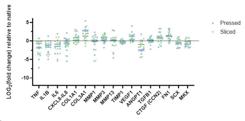
Discussion and conclusions: Different LHBT autograft preparation techniques do not induce substantial early alterations in gene expression or histopathological tendon integrity immediately after graft preparation. These preliminary findings suggest that LHBT patch processing does not markedly disrupt cellular signaling pathways or collagen architecture at baseline.



