
Deutscher Kongress für Orthopädie und Unfallchirurgie 2026 (DKOU 2026)
Mapping and classification of hip joint morphology: A large-scale CT-based analysis
Text
Objectives and questions: The McKibbin index, combining femoral and acetabular version, is a key determinant of hip stability and impingement risk. Aberrant combined version patterns may pose intraoperative challenges, yet their population distribution remains poorly defined. Moreover, the relationship between hip morphology, lower limb alignment, and pelvic parameters is not fully understood. The primary aim of this study is to comprehensively phenotype femoroacetabular anatomy in a large cohort and to evaluate correlations between the McKibbin index, hip morphology, lower limb alignment, and spinopelvic parameters.
Material and methods: We retrospectively analyzed 2,500 preoperative rotational CT planning datasets provided by Symbios S.A., all acquired using standardized protocols to ensure consistent image quality and geometry. A reproducible Python pipeline processed each case from raw imaging to structured measurement tables. Anatomies were segmented using TotalSegmentator. Predefined planning landmarks were combined with automated geometric landmarking. Sacral landmarking identified the center and surface normal of the superior sacral endplate, while tibial landmarking determined the tibial anatomical axis and proximal tibial plateau landmarks. Based on these landmarks, clinically relevant alignment and spinopelvic parameters were computed, including three-dimensional and coronal hip–knee–ankle angle, MPTA, LDFA, femoral neck length, femoral head position, pelvic incidence, and pelvic tilt. Hips were classified by femoral CCD angle into varus (<1 SD), neutral (±1 SD), and valgus (>1 SD). Group comparisons were performed using the Kruskal–Wallis test with Dunn’s post hoc correction for non-normally distributed data. Analyses were conducted using Python 3.9.6 and SciPy 1.13.1. Correlation analyses with lower limb parameters are ongoing.
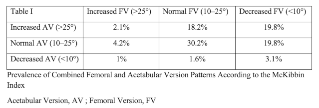
Results: All hips were classified according to CCD morphology: 6.8% varus (CCD < 124.45°), 84.4% neutral (124.45°–137.58°), and 8.9% valgus (> 137.58°). Femoral offset differed significantly across groups (p < 0.001), with valgus hips demonstrating significantly lower offset compared with both neutral and varus hips. Femoral anteversion also varied significantly (p = 0.004), with varus hips showing higher anteversion (18.69 ± 6.31°) than neutral (11.18 ± 11.24°) and valgus hips (5.71 ± 20.56°). In contrast, acetabular anteversion (overall mean 22.4° ± 7.5°) did not differ across CCD categories. Combined femoral and acetabular version patterns were heterogeneously distributed, with the majority falling within physiologic version ranges. The distribution of hip and acetabular anatomy using the McKibbin index is described in Table 1 [Tab. 1].
Discussion and conclusions: Large-scale CT phenotyping demonstrates that CCD morphology is associated with femoral offset and femoral anteversion, while acetabular anteversion remains independent. The McKibbin index shows a predominantly physiologic but heterogeneous distribution, providing a robust anatomical reference for preoperative planning.



